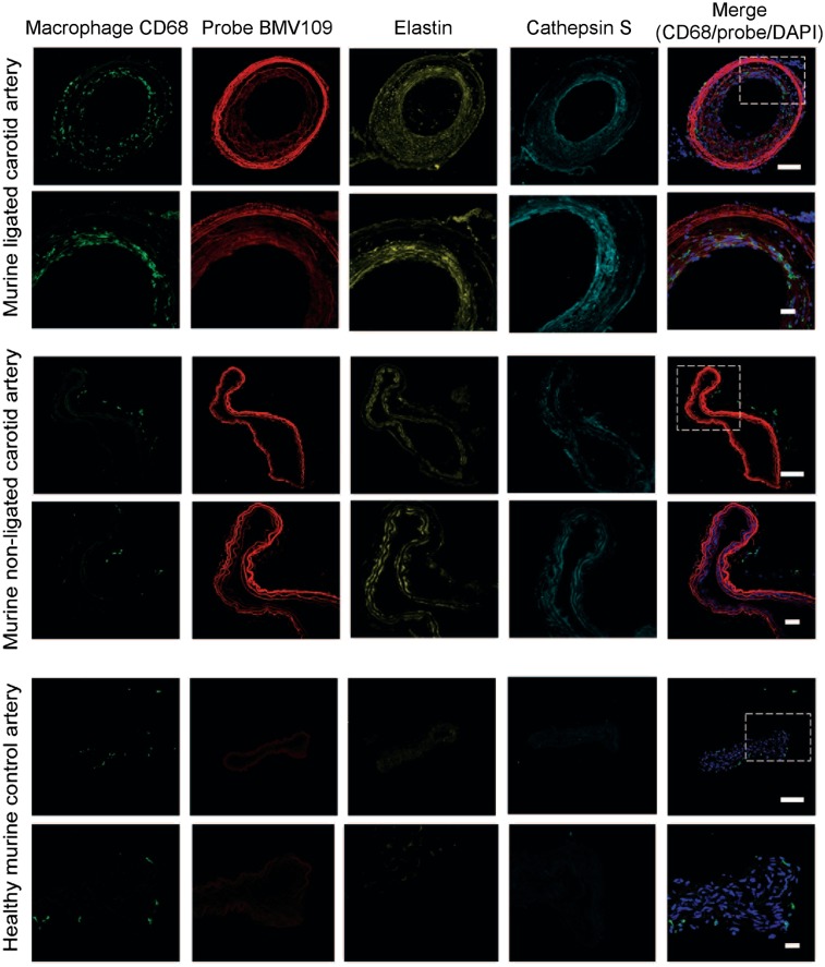
FIGURE 2.
Immunostaining of representative carotid arteries. Tissue cross-sections from ligated, nonligated, and control carotid arteries were labeled with optical probe BMV109 (red) and costained with macrophage activation marker CD68 (green), elastin (yellow), and cathepsin S (cyan). DAPI nuclear stain is shown in blue. Samples were tile-scanned at high resolution to generate full images for which scale bar represents 1 mm. White boxes on full images indicate region that higher magnification images were taken at 40×. Scale bars on zoom images are 10 μm.

