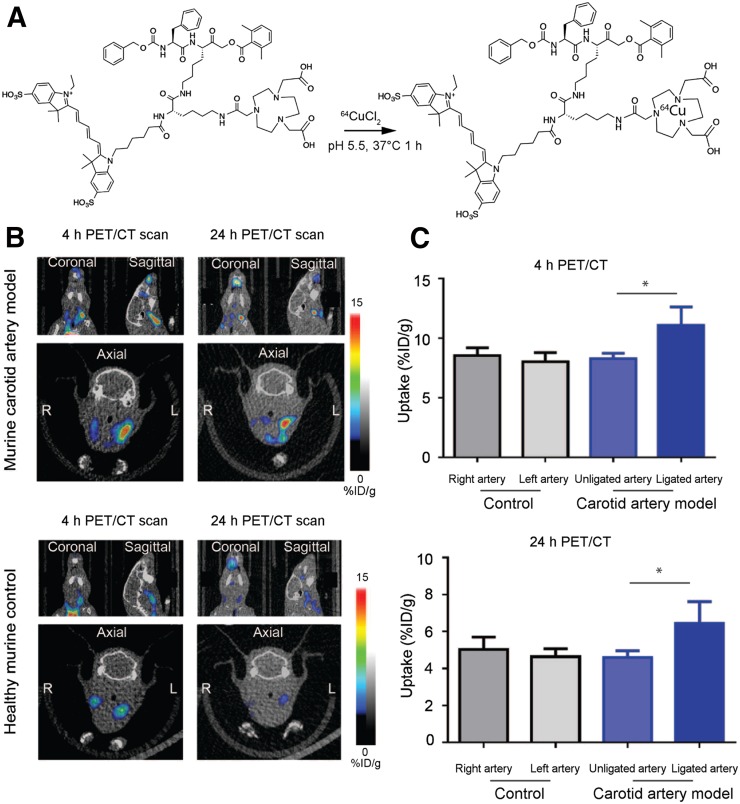
FIGURE 3.
Application of dual optical/PET imaging probe 64Cu-BMV101. (A) Structure and labeling conditions for dual optical/PET probe BMV101. (B) Noninvasive PET/CT scans of mice with and without ligated carotid arteries. Coronal (top left), sagittal (top right), and axial (bottom, showing left and right). Images are shown for representative diseased and healthy mice imaged at 4 and 24 h. (C) Quantification of 4- and 24-h PET/CT intensity from ligated, nonligated, and healthy carotid arteries of all mice. Error bars indicate mean + SEM, n = 3, *P < 0.05 by t test.

