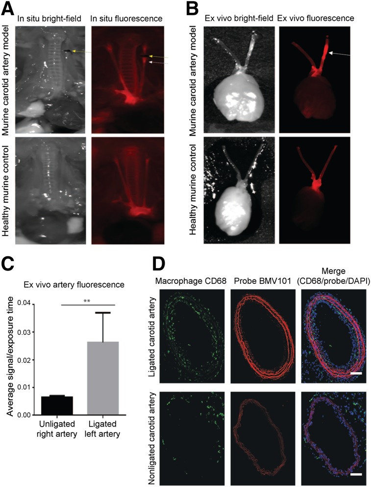
FIGURE 4.
In situ, ex vivo, and immunostaining of representative carotid arteries from 64Cu-BMV101–treated mice. (A) In situ fluorescence imaging of 64Cu-BMV101 in murine carotid arteries and control healthy mouse. (B) Ex vivo florescence imagining of diseased and healthy carotid arteries. (C) Quantitative analysis of ex vivo fluorescence showed significantly higher signal in left ligated carotid artery than nonligated carotid artery. n = 3, *P < 0.05 by t test. (D) Tissue sections from ligated and nonligated carotid arteries were labeled with optical probe 64Cu-BMV101 (red) and costained with macrophage activation marker CD68 (green). DAPI nuclear stain is shown in blue. Samples were tile-scanned at high resolution to generate full images for which scale bar represents 1 mm.

