FIGURE 5.
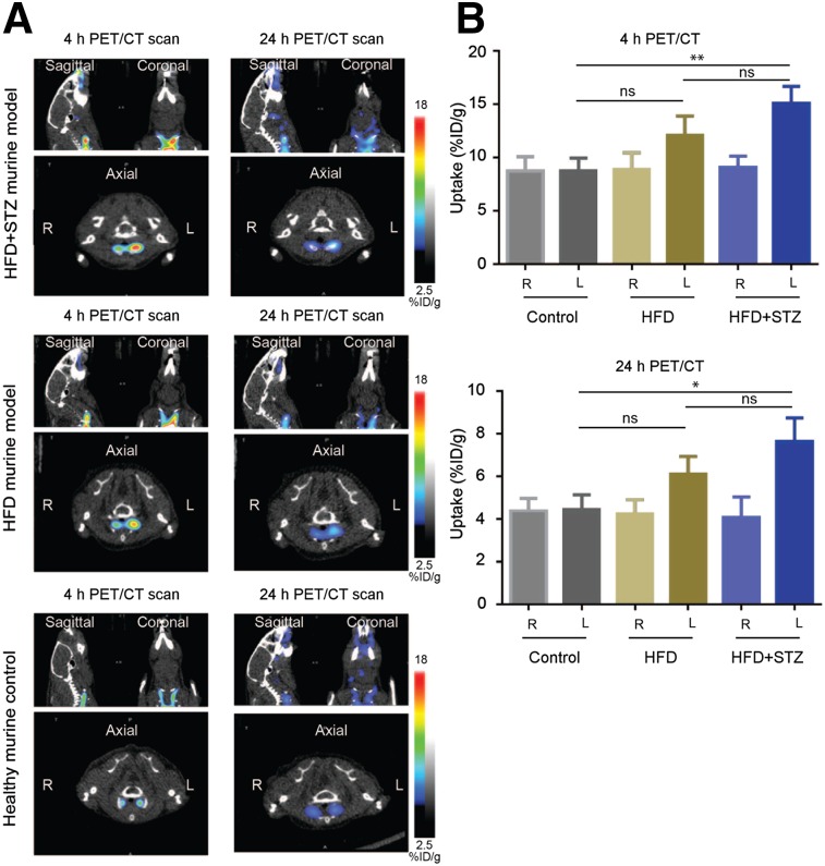
Comparison of dual optical/PET imaging probe 64Cu-BMV101 uptake in HFD + STZ model vs. HFD alone. (A) Noninvasive PET/CT scans of mice with and without ligated carotid arteries. Coronal (top right), sagittal (top left), and axial (bottom, showing left and right). Images are shown for representative HFD + STZ, HFD-alone, and healthy mice imaged at 4 and 24 h. (B) Quantification of 4- and 24-h PET/CT intensity from ligated HFD + STZ, HFD-alone, and nonligated healthy carotid arteries of all mice. Error bars indicate mean + SEM, n = 3, **P < 0.005, *P < 0.05 by t test.

