Figure 3.
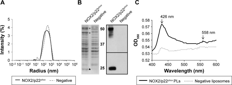
In vitro physicochemical characterization of NOX2/p22phox and negative liposomes.
Notes: (A) Particle size distribution of NOX2/p22phox and negative liposomes by DLS. (B) Coomassie blue staining of NOX2/p22phox and negative liposomes (The * and ▲ indicate the location of NOX2 and p22phox respectively) and Western blot analysis of NOX2/p22phox and negative liposomes using monoclonal antibodies against NOX2 and p22phox. (C) Dithionite-reduced minus-oxidized spectra of NOX2/p22phox and negative liposomes.
Abbreviations: DLS, dynamic light scattering; OD, optical density; PLs, proteoliposomes.

