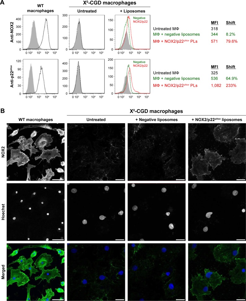
Figure 5.
Analysis of the membrane delivery of NOX2 and p22phox subunits in X0-CGD iPSC-derived macrophages after NOX2/p22phox liposome treatment.
Notes: (A) Flow cytometry analysis of NOX2 and p22phox expression using monoclonal antibodies in WT and untreated X0-CGD macrophages (black curve), and X0-CGD macrophages treated for 8 h with NOX2/p22phox (red curve) or negative (green curve) liposomes. Isotype controls are represented by gray-filled curves. MFIs were indicated for each condition, and the shift of fluorescence was calculated as the percentage of increased fluorescence compared to untreated macrophages. (B) Confocal microscopy images showing the staining of NOX2 subunit with 7D5 antibody and AF488-conjugated secondary antibody (green) in WT and X0-CGD macrophages treated for 8 h with NOX2/p22phox or negative liposomes. Nuclei were counterstained with Hoechst 33258 (blue); scale bars =20 µm. The same observations were obtained in at least two experiments.
Abbreviations: CGD, chronic granulomatous disease; iPSC, induced pluripotent stem cell; MFIs, mean fluorescence intensities; MΦ, macrophages; WT, wild type; X0-CGD, X0-linked CGD; XCGD, X-linked CGD.

