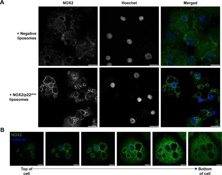
Figure 6.
Location of NOX2 in liposome-treated X0-CGD iPSC-derived macrophages after C. albicans phagocytosis.
Notes: (A) Confocal microscopy images showing the staining of NOX2 subunit with 7D5 antibody and AF488-conjugated secondary antibody (green) in X0-CGD macrophages treated for 8 h with NOX2/p22phox or negative liposomes and then for 4 h with C. albicans strain at an MOI of 3:1. (B) Z-stack images (top to bottom of cell) of an NOX2/p22phox liposome-treated X0-CGD macrophage incubated for 4 h with C. albicans strain at an MOI of 3:1. Nuclei were counterstained with Hoechst 33258 (blue); scale bars =20 µm in (A) and 10 µm in (B). The same observations were obtained in at least two experiments.
Abbreviations: C. albicans; Candida albicans, CGD, chronic granulomatous disease; iPSC, induced pluripotent stem cell; MOI, multiplicity of infection; X0-CGD, X0-linked CGD; XCGD, X-linked CGD.

