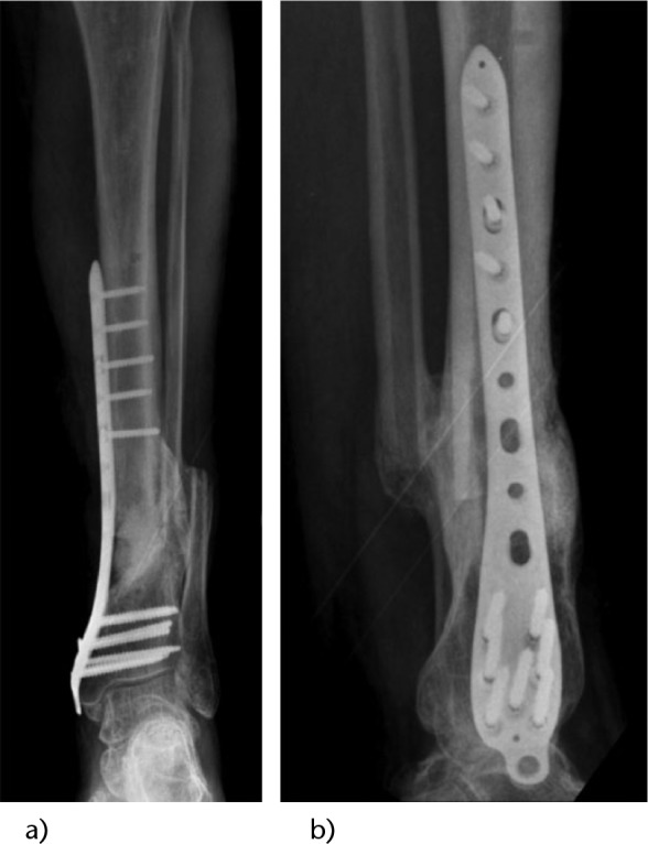
Fig. 3.

Radiographs taken nine months post-revision surgery, showing good incorporation of the graft and continuity of the tibia. a) Anteroposterior (AP) radiograph; b) lateral (LAT) radiograph.

Radiographs taken nine months post-revision surgery, showing good incorporation of the graft and continuity of the tibia. a) Anteroposterior (AP) radiograph; b) lateral (LAT) radiograph.