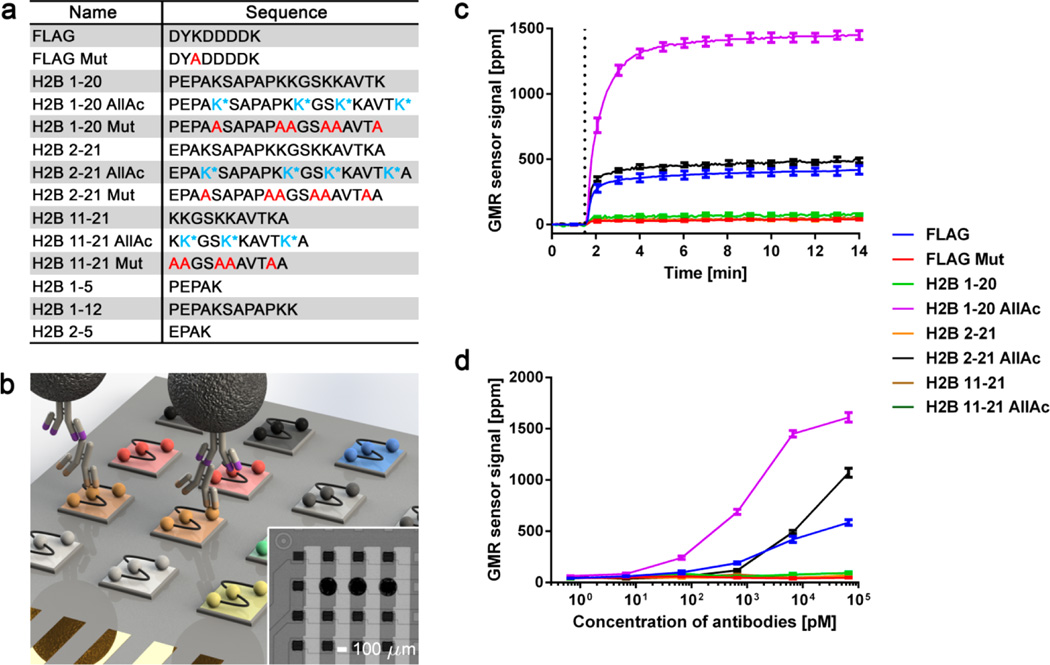
Figure 1.
Development and validation of GMR nanosensor peptide microarrays. (a) List of peptides used in GMR nanosensor microarray. Acetylated lysine (K*) and alanine substitution (A) are represented in blue and red, respectively. (b) Schematic of GMR nanosensor peptide microarray (not to scale). A panel of peptides (indicated by different colors) was spotted on a GMR nanosensor chip. Antibody-containing samples were used to probe the microarrays, combined with biotinylated secondary antibodies (illustrated with purple tips) and streptavidin-coated MNPs. The stray field from the bound MNPs was detected by the nanosensors underneath. Inset: 16 sensors from an 8 × 8 GMR nanosensor chip (peptides were spotted on 3 sensors in the second row from the top). (c) Real-time signals obtained from the GMR nanosensor peptide microarray probed with anti-FLAG and anti-K5Ac antibodies. MNPs were added to the microarray at ~1.5 min as indicated by the dotted line. (d) Titration curves of anti-FLAG and anti-K5Ac antibodies measured with the microarrays. Error bars represent standard deviations of 4 identical sensor signals.

