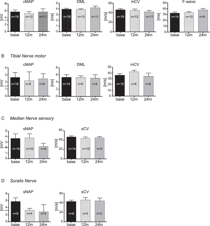
Figure 4.
Electrophysiological data. Median (A,C), tibial (B) and sural nerve (D) were analyzed at three time points: baseline, follow-ups after 12 months and after 24 months. Number of values differs indicated by (n=) in each bar as some patients reject single measurements; CMAP, compound motor action potential; DML, distal motor latency; mCV, motor conduction velocity; SNAP, sensory nerve action potential; sCV, sensory conduction velocity.

