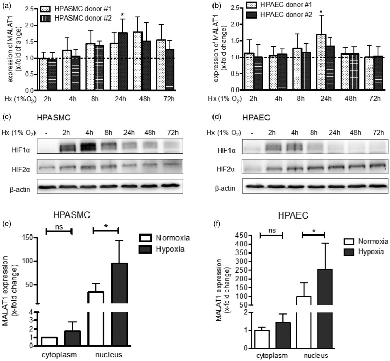
Figure 2.
Hypoxia increases the expression of MALAT1 in human pulmonary vascular cells. (a) Human pulmonary artery smooth muscle cells (HPASMC) from healthy donors were exposed to hypoxia (Hx, 1% O2) and, as shown by qPCR analysis, the levels of MALAT1 were found to be induced after 24 h of Hx. (b) Similarly, human pulmonary artery endothelial cells (HPAEC) from healthy donors showed higher expression levels of MALAT1 when exposed to Hx (1% O2) with a significant induction of MALAT1 after 24 h of Hx. (c) To confirm successful application of Hx in HPASMC, Western blot analysis was performed that demonstrated stabilization of HIF1α but not of HIF2α after exposure of cells to Hx. (d) Along that line, activation of HIF signalling in HPAEC was demonstrated by Western blot analysis. (e) The subcellular localization of MALAT1 in HPASMC was assessed. As demonstrated by qPCR analysis, MALAT1 is mainly localized in the nucleus when compared to the cytoplasmic fraction. Levels of MALAT1 could be further induced by Hx (24 h, 1% O2). (f) The same observation was made when the subcellular localization of MALAT1 was investigated in HPAEC. n = 4–6 for all experiments. Statistical analysis by one-way ANOVA with Tukey post hoc test (a, b, e, and f) and representative Western blot are shown (c and d) (*P < 0.05)

