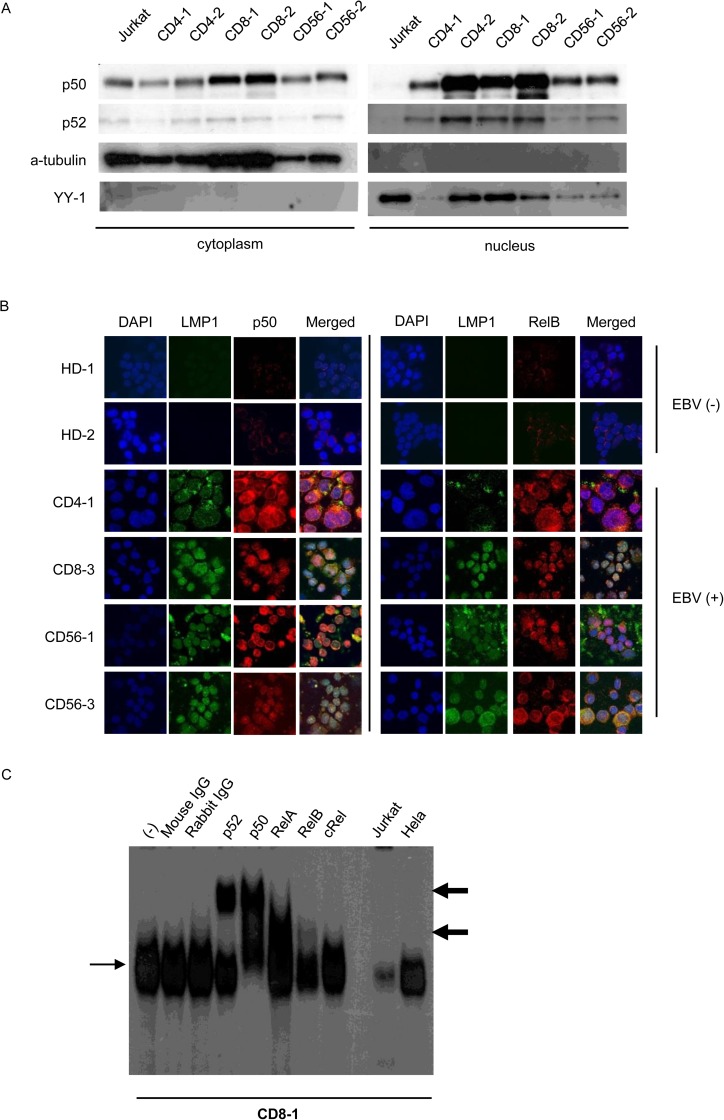
Fig 2. NF-κB activation in Epstein-Barr virus positive T or NK cells from patients with EBV-positive T or NK cell lymphoproliferative disorders.
(A) Western blotting for p50 and p52 protein localization in EBV-positive T or NK cells purified from patient peripheral blood mononuclear cells (PBMCs) using antibody-conjugated magnetic beads and immediately used in the assay. (B) Immunofluorescent staining for NF-κB protein localization in patient PBMCs. EBV-positive T or NK cells purified from patient PBMCs using antibody-conjugated magnetic beads were immediately subjected to immunofluorescent staining with anti-LMP1 and anti-p50 or anti-LMP1 and RelB antibodies. DAPI was used for nuclear staining. PBMCs from healthy donors were used as negative controls. Cells were analyzed via confocal microscopy. (C) Electrophoretic mobility shift assay of PBMCs from CD8-1. Nuclear extracts were obtained, incubated with KBF-1 containing the NF-κB binding sequence with or without indicated antibodies, and subjected to the assay. Thin, black arrows indicate KBF-1-binding NF-κB proteins. Thick, black arrows indicate supershifted bands. Nuclear extracts of Jurkat cells and HeLa cells treated with 10 ng/ml of TNF-α for 30 min were used as controls.

