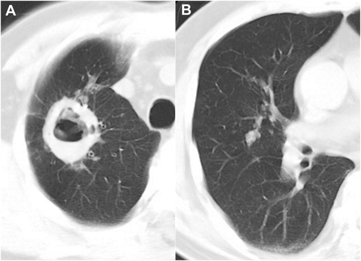
Fig 3. CT images of Mycobacterium tuberculosis infection in a 67-year-old man.
Chest CT axial images with a lung window settings (2.5-mm slice) were obtained at the levels of the superior vena cava and right middle lobar bronchus. The CT image show a cavity with a thick and irregular wall and multiple satellite tree-in-bud nodules in the right upper lobe (A). Note the multiple well-defined tree-in-bud nodules (>6 mm) in the right middle lobe (B).

