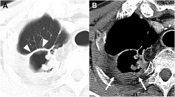
Fig 5. CT images of a Mycobacterium intracellulare pulmonary infection in a 77-year-old man.
Chest CT axial images were obtained at the level of the right bracheocephalic vein (2.5- mm slice). The lung window setting shows the thin and even thickness of the cavity (arrowheads) in the right upper lobe (A). The mediastinal window setting shows thick pleural thickening with proliferation of extra-pleural fat next to the cavity in the right upper lobe (arrows) (B).

