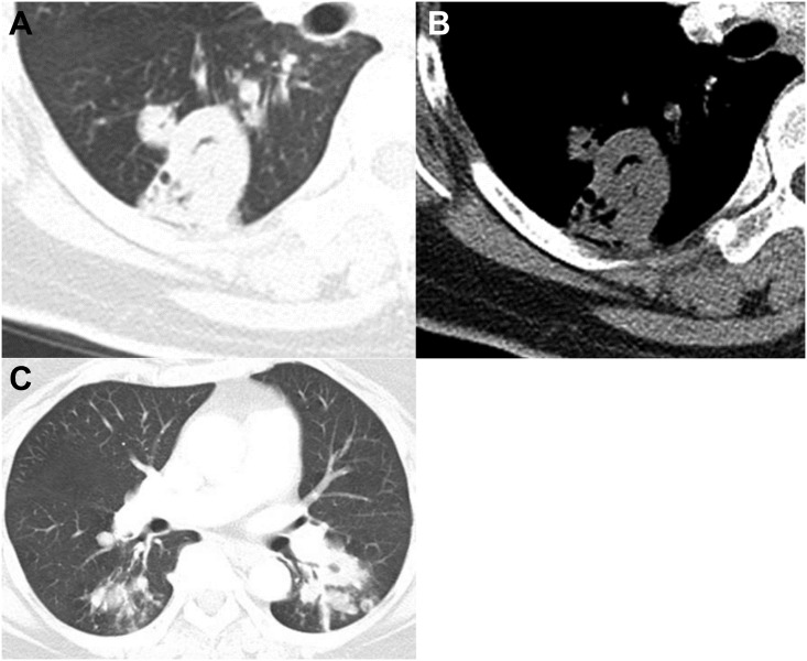
Fig 6. CT images of a Mycobacterium tuberculosis infection in a 73-year-old female.
Chest CT axial images (2.5 mm collimation) were obtained of the bronchus intermedius (A, B) and superior segmental bronchi of both lower lobes (C). The lung and mediastinal window settings show a thick-walled cavity in the superior segment of the right lower lobe (A) without marked pleural thickening next to the cavity (B). (C) Note the multiple satellite tree-in-bud nodules in the superior segment of both lower lobes (> 6 mm) and the macronodules in the left lower lobe (≥10mm).

