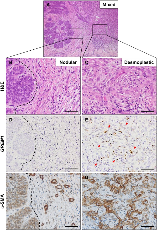
Fig 3. GREM1 expression in mixed type basal cell carcinoma.
A representative hematoxylin and eosin staining of a basal cell carcinoma (A) having both nodular (B) and desmoplastic (C) features. Fibroblasts around the nodular area (indicated by black dotted line) were negative for GREM1 (D) and α-smooth muscle actin (F), whereas those around the desmoplastic area were positive for both GREM1 (indicated by red arrow heads) (E) and α-SMA (G). RNA in situ hybridization for GREM1 and immunohistochemical analysis for α-SMA. Scale bar: 25 μm.

