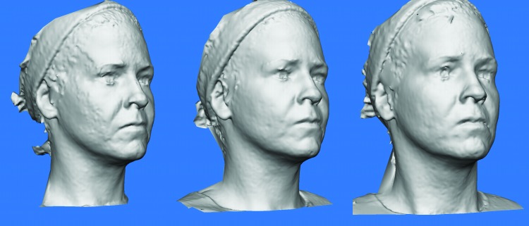
Figure 4.
Grey-scale 3D Vectra Photographic Evidence from Patient 05. A) Vectra 3D photo of patient 05 at baseline; Vectra 3D Photos of patient 05 at Week 2 (post-intradermal). Improved skin texture. Slight decrease in nasolabial folds, marionette lines. Increased distance between lateral canthus and inferior border of lateral brow; C) Vectra 3D Photos of patient 05 at Week 4 (post-intramuscular). Added improvement of glabellar and periorbital lines.

