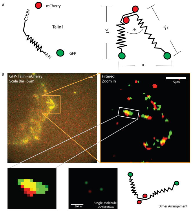
Figure 1.
Localization of the C and N termini of talin Dimers. (A) A schematic of the GFP-talin-mCherry construct (GTC). GFP is attached to the N-terminal of talin and mCherry to the C-terminal of Talin. The N–C terminal distance (termed stretch) is calculated by averaging the length of two strands of the dimer, that is, (y1 + y2)/2. The distance between the two N-termini is referred to as x. (B) The GTC construct was transfected into talin (−/−) cells. Images were processed by the previously described methods for subsequent single molecule identification and tracking. Lower panel represent the illustration of centroid-based localization of a single talin dimer.

