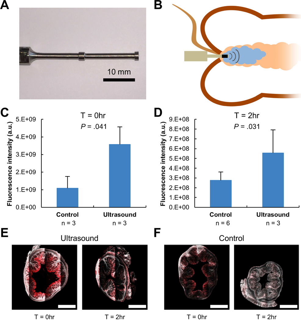
Figure 2. Ultrasound-Mediated GI Delivery of 3 kDa Dextran In Vivo.
(A) A custom-made 40 kHz ultrasound probe allowing for administration in the colon of mice. The protrusions enhance radial ultrasound activity. (B) Experimental setup depicting placement of an enema in the colon followed by ultrasound exposure with the custom probe shown in (A). Total fluorescent intensity (averages + 1SD) of the colon immediately after administration (C) of 3 kDa dextran tagged with Texas red with ultrasound or without ultrasound (Control) and two hours after administration (D). P-values determined by two-tailed Student’s t-tests. Representative multiphoton microscopy images of fixed murine colonic tissue after delivery of 3 kDa dextran tagged with Texas red with ultrasound (E) and without ultrasound (F) immediately and two hours after administration. The red channel and second harmonic are shown. The scale bars represent 500 µm.

