Figure 1.


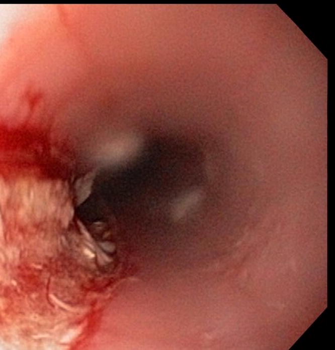
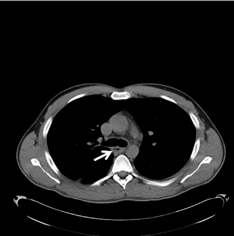
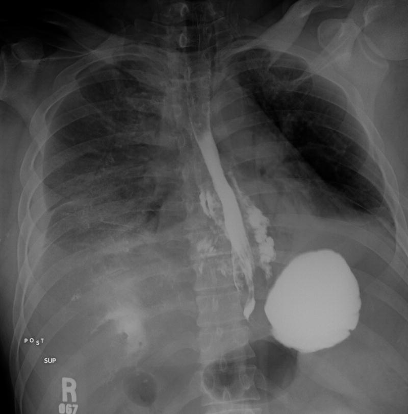
Examples of esophageal perforations in EoE patients. (A) Endoscopic view of a deep mucosal rent concerning for an esophageal perforation. (B) Noncontrasted chest CT scan in a different patient demonstrating diffuse esophageal wall thickening with associated paraesophageal stranding. A small amount of free mediastinal air can be seen (arrow), as well as paraesophageal fluid suggestive of phlegmon vs. abscess, suggesting a transmural perforation. (C) Barium swallow in a different patient with free extravasation of contrast into the mediastinum consistent with a transmural perforation.
