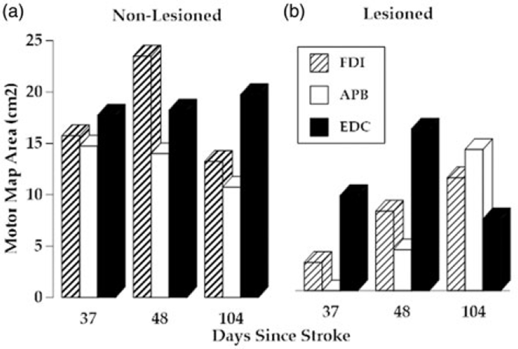
Figure 4. Motor maps.
(a) Non-lesioned hemisphere motor map area for the first dorsal interosseous muscle (blue columns), the adductor pollicis brevis (red columns) and extensor digitorum communis (green columns). Data collection occurred on the same day as the clinical and kinematic measures from Figures 2 and 3. (b) Lesioned hemisphere motor map area for the same muscles collected on same days.

