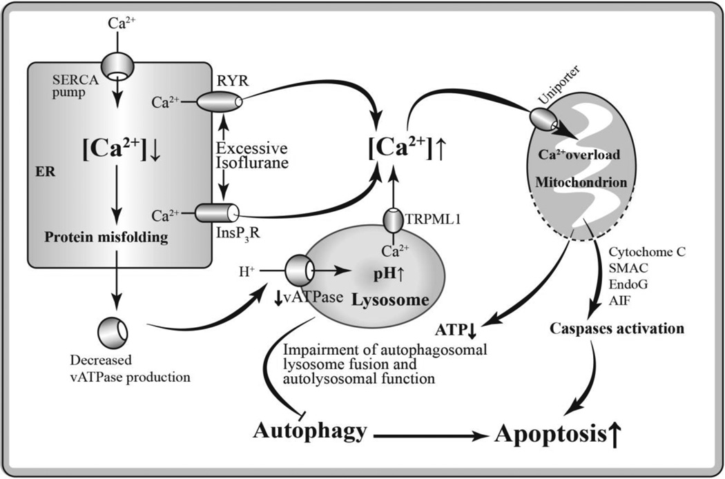
Fig. 2.
Hypothetical pathways of anesthetic neurotoxicity via impaired autophagy secondary to intracellular Ca2+ dysregulation. A prolonged exposure of GAs such as isoflurane at high concentration induces excessive Ca2+ release from the ER into the cytoplasm by over-activation of InsP3Rs and/or ryanodine receptor (RYR). The decreased Ca2+ level in ER causes accumulation of misfolded protein including vATPase, resulting in elevated lysosomal pH value and impaired autolysosomal function. Abnormally increased cytosolic Ca2+ leads to mitochondrial Ca2+ overload, and resulting in uncoupling of oxidative phosphorylation and release of catabolic hydrolases such as cytochrome c, apoptosis inducing factor (AIF), endonuclease G (EndoG) and secondary mitochondria-derived activator of caspase (SMAC), and result in apoptosis. In addition, the mitochondrial Ca2+ overload may induce decreased ATP production and inhibited ATP-dependent autophagic cytoprotection, thus promoting apoptosis.

