
Figure 2.
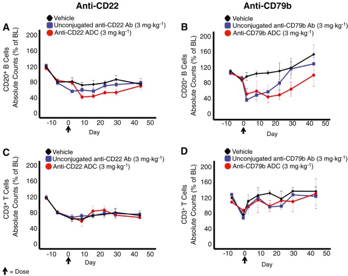
Depletion of CD20+ B cells following administration of anti‐CD22 or anti‐CD79b ADCs. Peripheral blood CD20+ B cells (panels A and B) or CD3+ T‐cells (panels C and D) in animals administered either anti‐CD22 (panels A and C) or anti‐CD79b (panels B and D) ADCs (n = 4); respective unconjugated antibodies (Ab) (n = 4) or vehicle (n = 4) over time are presented. Data presented are group mean absolute counts expressed as a percentage of baseline (% of BL) at each time point. Baseline was calculated as the average value obtained at two pre‐dose time points (shown). Error bars represent SEM. The arrow denotes administration of the test substances.