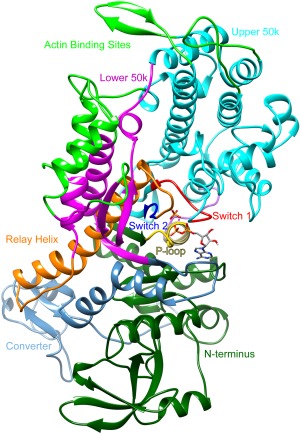
Figure 1.

Structure of dictyostelium discoideum myosin S1 head. Functional regions highlighted in different colors: Upper 50k, cyan; Lower 50k, magenta; actin binding sites, bright green; P‐loop, yellow; Switch 1, red; Switch 2, blue; Relay helix, orange; Converter, light blue; N‐terminus, dark green.
