. 2017 Mar 15;58(3):570–580. doi: 10.3349/ymj.2017.58.3.570

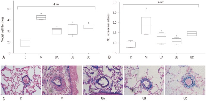
Fig. 5. Pathologic findings in MCT-induced PAH rats according to dose of hUCB-MSCs at week 4. (A) Low-dose of hUCB-MSCs also effectively decreased pulmonary arteriole medial wall thickening. (B) The number of intra-acinar arteries was decreased in UA and UB groups. (C) Low-dose hUCB-MSCs also attenuated pulmonary pathological changes in MCT-induced PAH rat (Victoria blue staining, ×400). *p<0.05, compared with C, p<0.05, compared with M. No, number; C, control group; M, monocrotaline group; UA, 3×10⁶ cells/rat; UB, 1.5×10⁶ cells/rat; UC, 3×10⁵ cells/rat; MCT, monocrotaline; PAH, pulmonary arterial hypertension; hUCB-MSCs, human umbilical cord blood derived mesenchymal stem cells.

Fig. 5