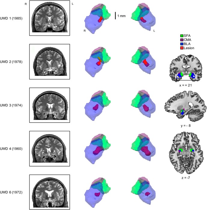
Figure 1.
Location and size of the BLA damage. Coronal view of T2-weighted MRIs (left) and a 3D reconstruction (middle) of the lesion for the five individuals with UWD with birth year indicated. Reconstruction of the AMG subnuclei was based on the cytoarchitectonic probability maps from Amunts et al., (2005) in Eickhoff et al., (2005). Black rectangle indicates viewpoint for the 3D reconstruction (right).

