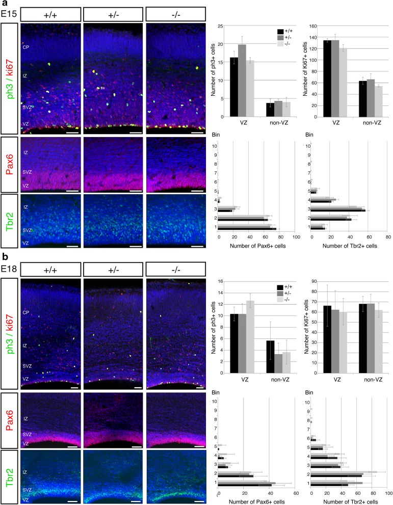
Fig. 2.
Neurogenesis is not altered in Kiaa0319 mutants. a Immunostaining of Kiaa0319 +/+, +/− and −/− embryos at E15.5. There are no differences between the three genotypes in the numbers or distribution of dividing (pH3 + , green) or cycling (Ki67 + , red) cells along the cortical wall (upper panels). Staining for Pax6 (red, middle panels) and Tbr2 (green, lower panels) also shows no changes in Kiaa0319 deficient animals. Nuclei were stained with DAPI (blue). Quantifications of number of cells per 100-μm wide sections in the ventricular zone and rest of cortical wall (upper panels) or per bin (middle and lower panels) are shown on the right. b Immunostaining of +/+, +/− and −/− embryos at E18.5. The number of dividing (pH3 + , green) or cycling (Ki67 + , red) cells and their distribution, quantified as number of cells per 100-μm wide section in the ventricular zone and the rest of the cortical wall, shows no variation between +/+, +/− and −/− animals (upper panels). Immunostaining against Pax6 (red, middle panels) and Tbr2 (green, lower panels) shows similar numbers and distribution of positive cells in all three conditions. Nuclei were stained with DAPI (blue). Quantifications are shown on the right. VZ ventricular zone, SVZ subventricular zone, IZ intermediate zone, CP cortical plate. Scale bars 75 μm

