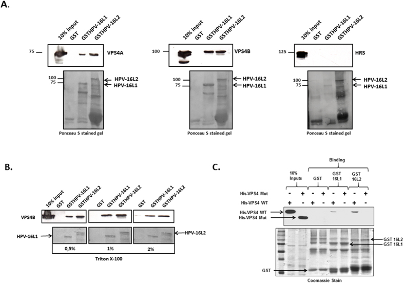
Figure 2. The ESCRT component VPS4 interacts with HPV-16 L1 and L2.
Panel A. HEK293 cells were transfected with VPS4AGFP, VPS4BDsRED or HRSGFP expressing plasmids. 24 h after transfection the cells were lysed and extracts incubated with purified GSTHPV-16L1, GSTHPV-16L2 or GST alone as a control, for 2 h at room temperature. After extensive washing the bound protein were analyzed by Western blotting against VPS4A, VPS4B and HRS (upper panel). The lower panel shows the Ponceau stain of the nitrocellulose membrane. Panel B. HEK293 cells were transfected with VPS4BDsRED, and after 24 h the cells were lysed and extracts incubated with GSTHPV-16L1, GSTHPV-16L2 or GST alone as a control. The beads were then washed with different concentrations of Triton-X-100 (0, 5%, 1%, 2%) in PBS and bound proteins were analyzed by western blotting using anti VPS4B antibody. Panel C. The HPV-16 L1 and L2 GST fusion proteins were purified and used in interaction assays with purified His-tagged VPS4B wild type (WT) and VPS4B delta MIT mutant (Mut) proteins. After extensive washing bound VPS4 was detected by western blotting and the results shown in the upper panel. The left two lanes show the VPS4 protein inputs. The lower panel shows the Coomassie stain of the fusion proteins used in the binding assays.

