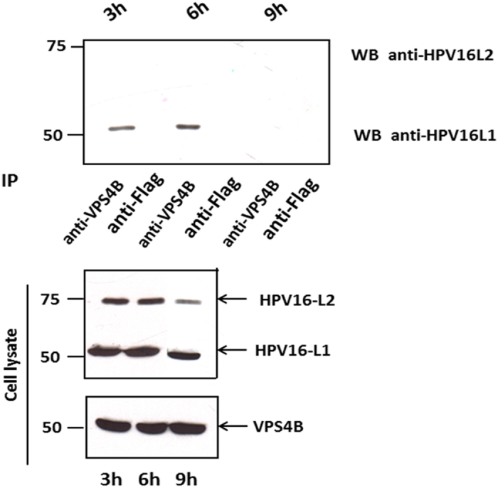
Figure 3. VPS4B interacts with HPV-16 L1 during viral infection.

HaCaT cells were infected with HPV-16 PsVs for 1 h at 4 °C. Cells were then washed with PBS and incubated at 37 °C. At 3 h, 6 h, 9 h post-infection the cells were harvested and cell extracts incubated with anti-VPS4B or rabbit anti-Flag antibody (control) overnight at 4 °C. The immunoprecipitates were then incubated with protein A conjugated sepharose beads for 1 h at room temperature. Any co-immunoprecipitating L1 and L2 proteins were then detected by western blotting using anti-L1 and anti-L2 antibodies (top panel). The lower panel shows the protein inputs for VPS4B, L1 and L2.
