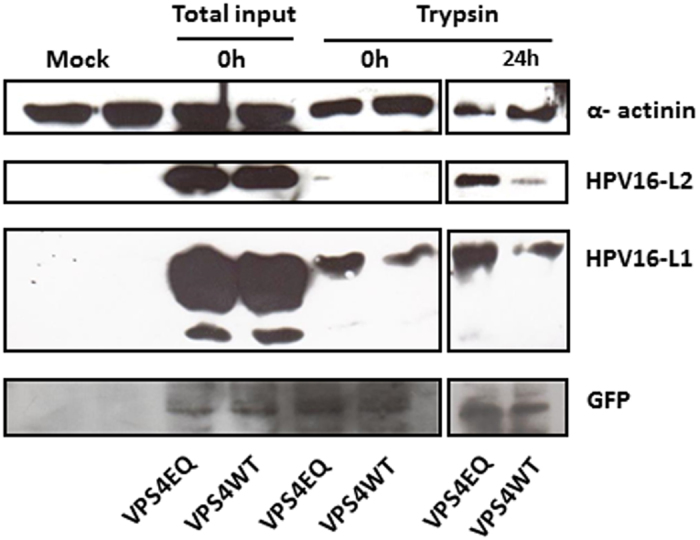
Figure 7. HPV-16 L1 and L2 appear more stable in mutant VPS4EQ expressing cells.

The 293-VPS4WT-GFP and 293-VPS4EQ-GFP cells were induced with 1 μM ponA and after 24 h were infected with HPV-16 PsVs for 1 h at 4 °C. Cells were then washed and incubated at 37 °C for 24 h. The cells were harvested at the different times and L1 and L2 levels analysed by western blotting. The extracts are as follows: the zero time-points corresponds to the total cell lysate collected after 1 h infection (total input virus) or the lysate from cells which were first treated for 15 minutes with trypsin after 1 h infection (to detect internalized virus); the cells harvested at 24 h were also treated with trypsin 15 mins prior to harvest to remove any residual non-internalized virus and therefore represent the residual internalized L1 and L2. Alpha-actinin was used as a loading control and the induced VPS4 proteins were detected using western blotting against GFP. Note the apparent increase in L1 and L2 levels in the mutant VPS4EQ expressing cells.
