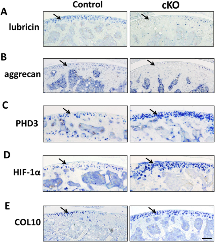
Figure 4. Expression of markers of articular cartilage progenitors and chondrocyte differentiation in control and cKO mice.
Immunohistochemistry analysis of markers in tibial articular cartilage of 12 week old control and cKO mice. (A,B) Expression of SZ progenitor markers, lubricin and aggrecan, in articular cartilage. (C) PHD3 expression in control and cKO articular cartilage. (D) HIF-1α expression in control and cKO articular cartilage. (E) Expression of the chondrocyte hypertrophy marker COL10 in control and cKO articular cartilage. Blue is positive staining. Arrows show positive stains of each protein in the articular cartilage. Bar = 50 μm.

