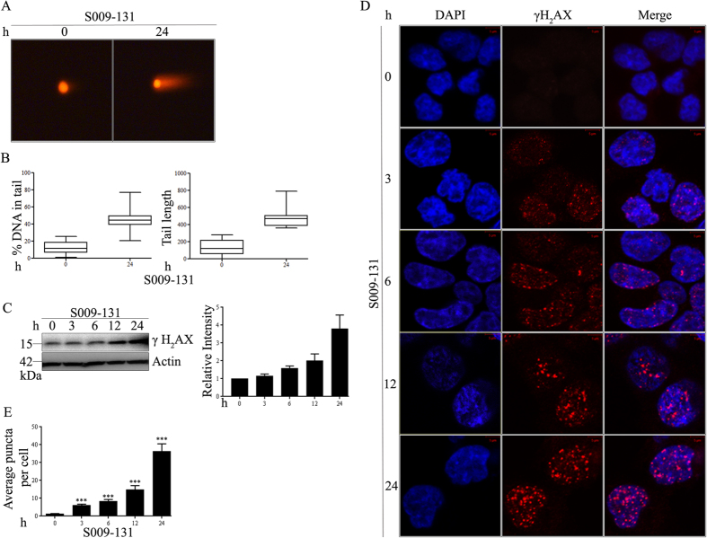
Figure 1. S009-131 induced DNA damage in HCT116 cells.
(A) Cells were treated with the molecule at 7.5 μM for 24 h and comet assay was done to determine the extent of DNA damage. (B) Tail length and percentage of DNA in tail was measured (minimum 28 cells were analysed for each treatment condition) by Image J and represented with box-and-whisker plot. (C) HCT116 cells were incubated with S009-131 for different time period. Cell lysates were then subjected to immunoblotting using anti-γ-H2AX antibody and analysed densitometrically. (D) Representative confocal images to show the γ-H2AX puncta at different time intervals on S009-131 treatment. (E) Bar graph showing average puncta per cell at different time interval (minimum 24 cells were considered in each group).

