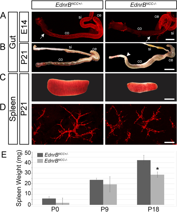
Figure 1. Embryonic and postnatal hindgut and spleens of EdnrBNCC+/− and EdnrBNCC−/− animals.
(A) tdTomato visualization of NCC in the small intestine and colon shows delayed colonization of the colon in EdnrBNCC−/− animals compared to EdnrBNCC+/−, with the migratory wavefront of NCC marked by white arrows at E14.5. (B) Aganglionosis in the distal colon of EdnrBNCC−/− animals at P21 causes functional obstruction (marked by white arrowhead). Normal, pelleted stool is seen in the distal EdnrBNCC+/− colon. (C) Reduced splenic size of EdnrBNCC−/− compared to EdnrBNCC+/− animals at P21. (D) tdTomato expressing NCC in EdnrBNCC+/− and EdnrBNCC−/− spleens at P21. (E) Spleens were harvested from EdnrBNCC+/− and EdnrBNCC−/− animals at P0, P9 and P18 and weighed. (*p < 0.05). Scale bars: A 400 μm, B 1 cm, C 4 mm and D 100 μm.

