Figure 3.
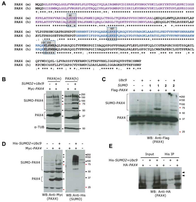
Mouse and human PAX4 proteins are SUMOylated. (A) Alignment of mouse PAX4 (PAX4 (m) UniProtKB P32115) and human PAX4 (PAX4 (h) UniProtKB O43316) protein sequences using Clustal Omega. PD is indicated in purple and HD in blue. The predicted SUMOylation sites are indicated by grey squares. (B) Western blot of cellular extracts from 293T cells transfected with Myc-tagged mouse Pax4 (left panels) or human PAX4 (right panels) alone or in combination with SUMO2 and Ubc9. Co-transfection of PAX4 with SUMO2 and Ubc9 results in the detection of SUMOylated PAX4. α-Tubulin was used as loading control (lower panels). Interestingly, human PAX4 SUMOylation is stronger when compared with mouse PAX4. In both cases, the co-expression of SUMO2 and Ubc9 seems to increase the stability of PAX4 protein, as indicated by the stronger band for unSUMOylated PAX4. (C) Western blot analysis of 293T cells transfected with Flag-tagged PAX4 alone or in combination with Ubc9, SUMO1 or SUMO2, shows human PAX4 SUMOylation by both SUMO1 and SUMO2 only when Ubc9 is co-expressed. (D) Analysis of extracts of 293T cells untransfected or transfected with Myc-tagged PAX4 alone or in combination with His-Tagged SUMO2 and Ubc9, using anti-Myc (left panel) or anti-His (right panel) antibodies, revealed the specificity of the SUMOylated bands. The lower band ~20 kD in the anti-His blot corresponds to free SUMO. (E) Pull down using His-Trap matrix of extracts from 293T cells transfected with Ha-tagged PAX4 alone or in combination with His-tagged SUMO2 and Ubc9 showing the specific pull down of SUMOylated PAX4, indicated by arrows.

