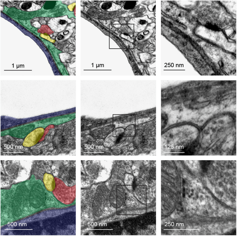
Figure 9.
Presence of synapses abutting astrocyte endfeet. Representative transmission electron microscopy images of cortical astrocyte endfeet surrounding capillaries of a 2-month-old C57BL6 mouse. Enlarged views of the squared areas show details of the synapses abutting astrocyte endfeet. On the left image, astrocyte endfeet are colored in green, endothelial cells in blue, pre-synapses in red and post-synapses in yellow.

