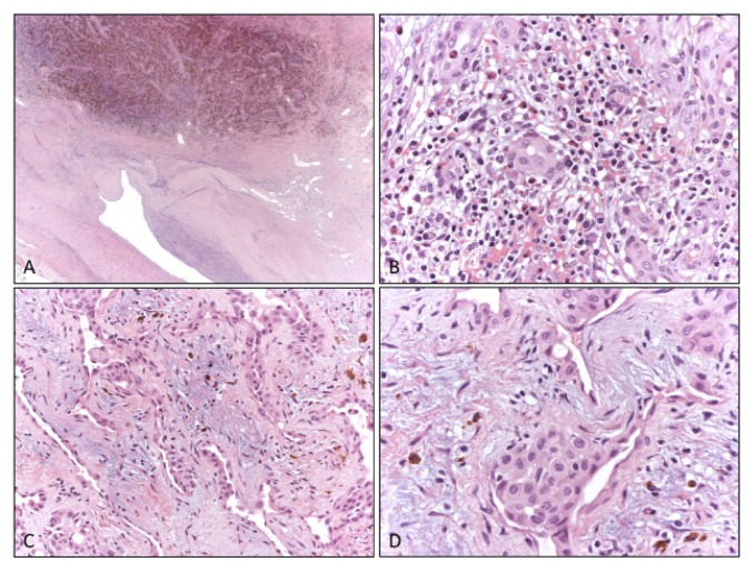
Figure 4.
Histologic appearance. At low magnification the arterial lumen was compressed and dislocated by a thickening of the tunica media of the vessel wall, that is replaced by fibrotic tissue with conspicuous hemosiderin deposits and vessels proliferation (A). At high magnification a heavy chronic inflammatory infiltrate including numerous eosinophils was present (B) and small irregular vessels proliferation (C) with prominent endothelial cells were also evident (D).

