
Figure 7.
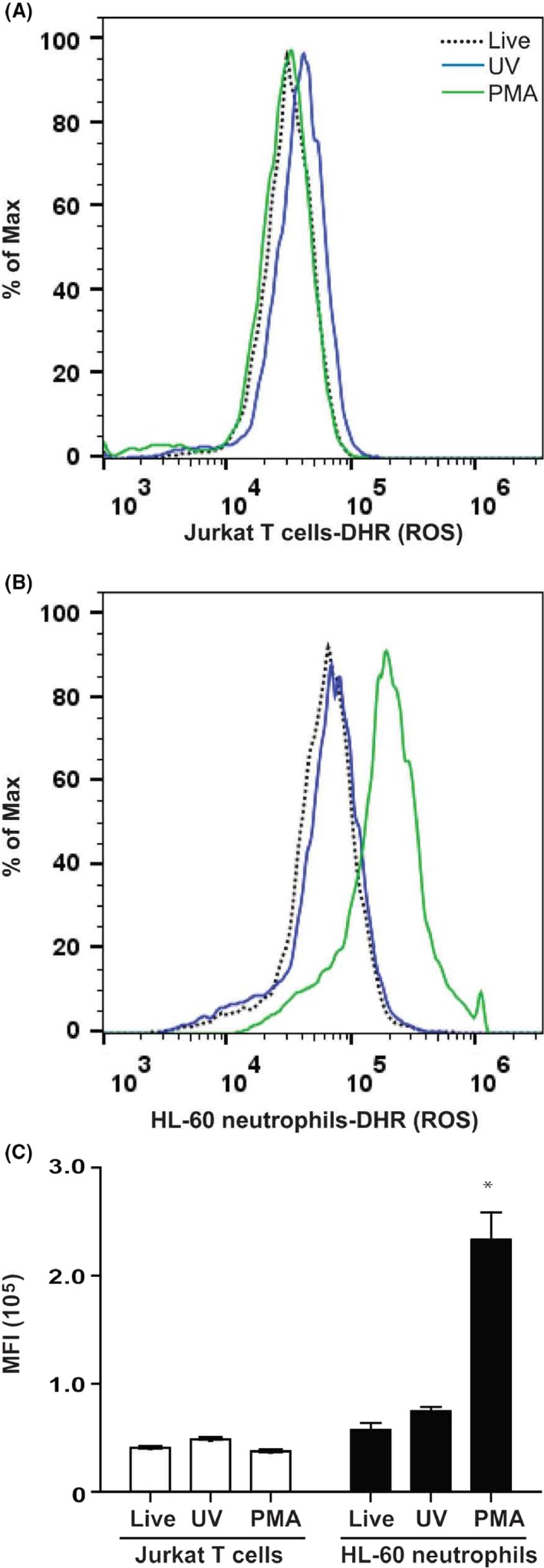
Flow cytometry correctly detects the presence of ROS inside the cells. Jurkat T cells and HL‐60 neutrophils were treated with 25 nmol/L PMA or irradiated with UV light. Fluorescence intensity was measure after 1‐h post incubation with DHR123. (A) Flow cytometry detects no ROS signal in Jurkat T cells after 60 min. (B) Flow cytometry detects a large amount of ROS in >75% of HL‐60 neutrophils compared to live cells or UV irradiated cells. (C) Mean fluorescence intensity shows that HL‐60 neutrophils treated with PMA generate large amounts of ROS compared to T cells and all other conditions (*,P < 0.05). N = 3; Experiments were conducted in three different days; each sample was analyzed in duplicate wells. Representative tracings are shown in A and B. Data were analyzed by two‐way ANOVA with Bonferroni post‐tests. ROS, reactive oxygen species.