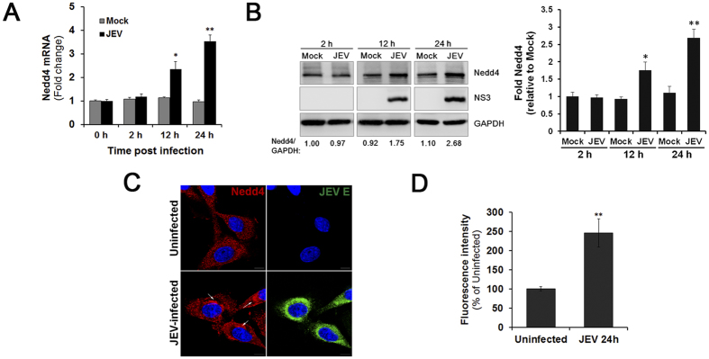
Figure 1. Nedd4 expression is up-regulated in SK-N-SH cells during JEV infection.
SK-N-SH cells were infected with JEV at an MOI of 1.0. The cells were collected at different time points. (A) Nedd4 mRNA levels were determined by RT-qPCR at different time points p.i. (B) The protein levels of Nedd4 and JEV NS3 were examined in mock- and JEV-infected SK-N-SH cells via Western blot using the indicated antibodies. GAPDH served as a control for equal sample loading. The level of Nedd4 was quantitated by densitometric analysis using ImageJ software and normalized to GAPDH. (C) Localization of Nedd4. SK-N-SH cells were fixed at 24 h p.i. and were subjected to immunofluorescence to detect JEV E (green) and Nedd4 (red). The nuclei were stained with DAPI (blue). Images were obtained using a confocal microscope (Scale bar = 10 μm). (D) The fluorescence intensity of Nedd4 was processed and quantified using ImageJ software. The data are shown as the mean ± SD of three independent experiments. *P < 0.05; **P < 0.01.

