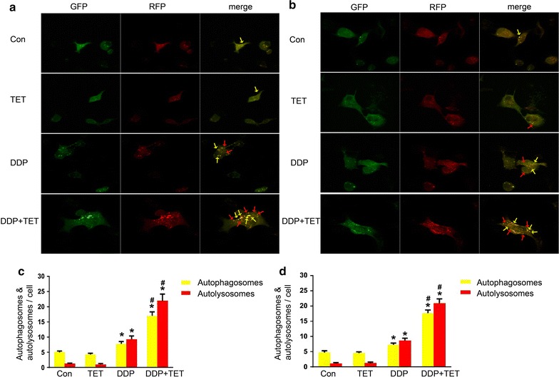
Fig. 9.

Autophagic flux assay of cisplatin-resistant A549 cells (a) and cisplatin-sensitive A549 cells (b) (×400). The concentrations of DDP in a, b were 60 and 25 µM, respectively. The concentration of TET was 0.25 µg/ml. The time of the drug treatments was 12 h. Yellow arrows point to autophagosomes and red arrows point to autolysosomes. c, d are the statistical analyses of a, b. *p < 0.05 versus the control cells, #p < 0.05 versus the DDP treatment (n = 3)
