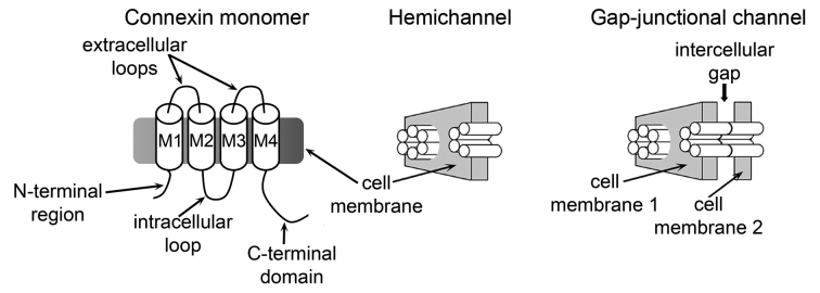
Figure 1.
Connexin channels and hemichannels. Schematic representation of a connexin subunit (monomer), a hemichannel (hexamer), and a gap-junction channel (dodecamer). M1 to M4: transmembrane helices. Each monomer is depicted as a cylinder in the hemichannel and the gap-junction channel. This figure is reproduced from one originally published in the J Biol Chem [30], and is reproduced with permission from the American Society for Biochemistry and Molecular Biology.

