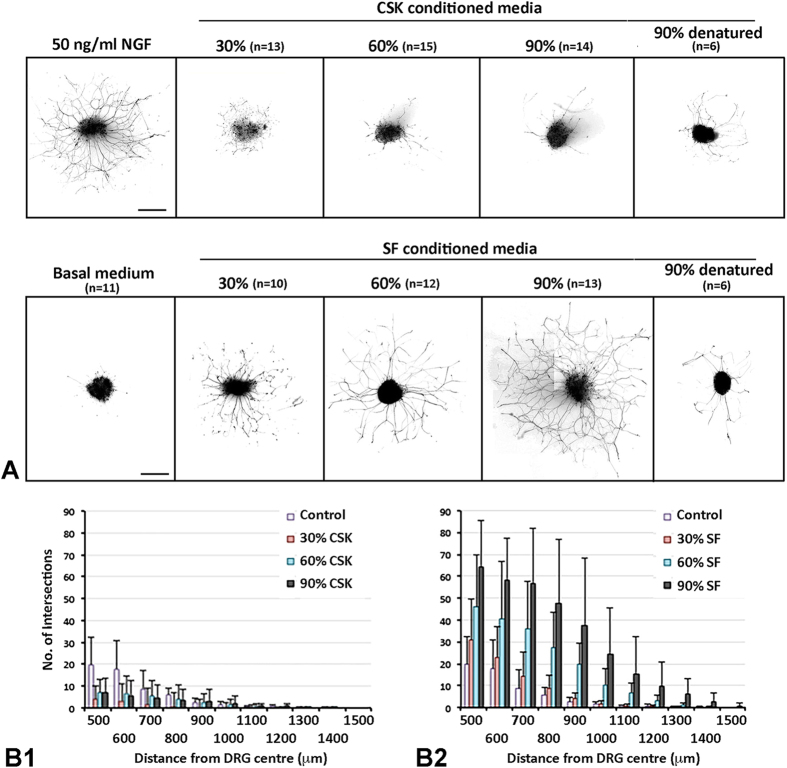
Figure 3. Neurite growth profile in DRG cultured with conditioned media from CSKs versus SFs of same donor.
(A) Processed images showing representative TuJ1-stained neurite growth from DRGs cultured in different dosages of conditioned media from CSKs or SFs or in control media at 72 hours. n - number of DRG in experiment. Scale bars: 500 μm. (B) Histograms showing the neurite extension pattern using concentric circle intersection counting method. (B1) CSK conditioned media; (B2) SF conditioned media. Data are presented as mean and standard deviation.

