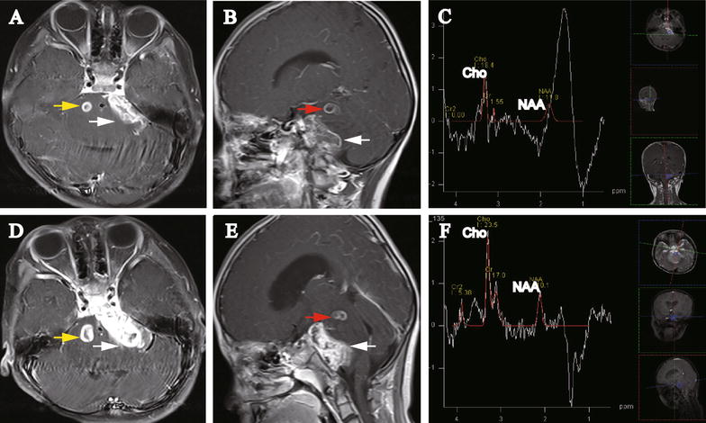
Fig. 2.

Images of repeated MRI on Dec 11, 2015 and Jan 12, 2016 for the 5-year-old girl with multiple posterior fossa lesions. A Axial gadolinium-enhanced MRI (Dec 11, 2015) displays the occurrence of a new lesion (0.8 cm × 0.9 cm, yellow arrow). The left CPA lesion is enlarged and extended along the cistern of the CPA (3.1 cm × 1.2 cm, white arrow). B Sagittal gadolinium-enhanced MRI (Dec 11, 2015) shows that the CPA lesion was enlarged and extended along the cistern of CPA (white arrow) and the upper left pontine lesion was enlarged (red arrow). C MRS (Dec 11, 2015) shows increased Cho and decreased NAA expression. The Cho:NAA ratio was also significantly increased. D Axial gadolinium-enhanced MRI (Jan 12, 2016) displays that both the CPA lesion (white arrow) and the new lesion in the right side of the pons are enlarged (1.1 cm × 1.3 cm, yellow arrow). The CPA lesion is extended along the cistern of the CPA. E Sagittal gadolinium-enhanced MRI (Jan 12, 2016) shows that both the CPA lesion (white arrow) and upper left pontine lesion (red arrow) were enlarged. F MRS (Jan 12, 2016) shows further increased Cho and decreased NAA expression. The Cho:NAA ratio was 2.33
