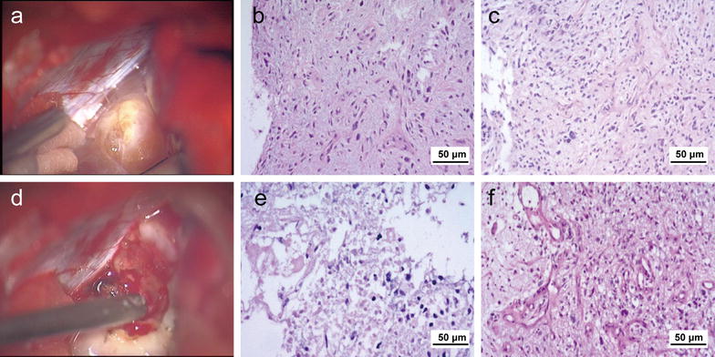
Fig. 3.

Histopathologic examination of the CPA lesion with hematoxylin–eosin staining. a Macroscopically, the outer layer of CPA lesion is rubbery and nodule-like. b Under a microscope, the outer layer of the CPA lesion displays moderate mitoses and necrosis. c Under a microscope, the outer layer of the CPA lesion displays moderate vascularization. d Macroscopically, the internal portion of the lesion is softer and has a higher blood supply than the outer layer. e Under a microscope, the internal portion of the CPA lesion displays obvious mitoses and necrosis. f Under a microscope, the internal portion of the CPA lesion displays hyper-vascularization
