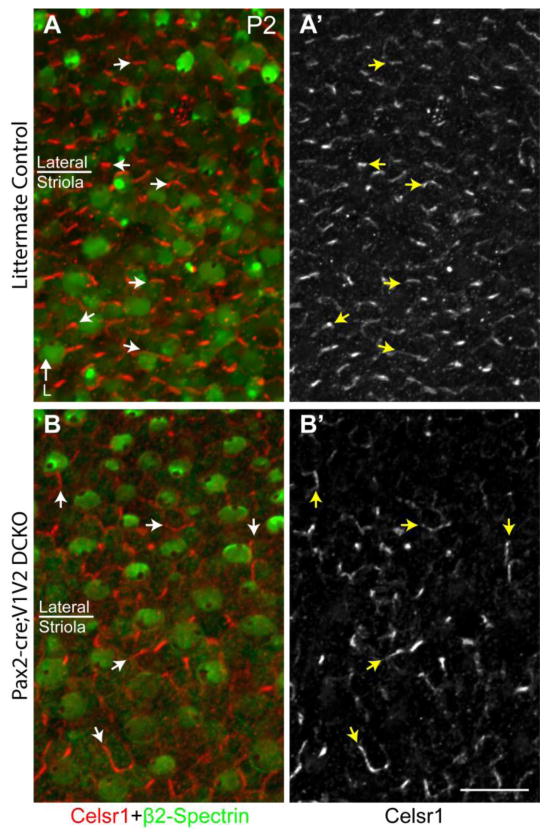
Figure 7. The polarized distribution of Celsr1 persists in the absence of Vangl1 and Vangl2.
(A) Celsr1 (red) and β2-Spectrin (green) labeling of the utricle from P2 littermate control mouse in a region spanning the boundary between the striolar and lateral region. Celsr1 is localized to SC:SC boundaries and organized along an axis parallel to stereociliary bundle orientation. (A′) Grayscale image of Celsr1 immunoflourescent labeling from ‘A’. (B) Celsr1 and β2-Spectrin labeling of the utricle from P2 Pax2-Cre; Vangl1; Vangl2 CKO mouse. Stereociliary bundles are misoriented relative to each other in all regions consistent with the loss of PCP signaling throughout the maculae. Bundle orientation can be inferred from the position of the fonticulus in the β2-Spectrin labeled hair cell apical surface. (B′) Grayscale image of Celsr1 immunoflourescent labeling from ‘A’. Despite the loss of Vangl1 and Vangl2, Celsr1 is still found to be asymmetrically localized at SC:SC junctions but no longer appears coordinated with stereociliary bundle polarity. Arrowheads illustrate examples of Celsr1 at HC:SC boundaries, arrows illustrate examples of Celsr1 at SC:SC boundaries. Anatomical reference arrow points along the Lateral (L) axis of the inner ear. Scale bar is 20μm.

