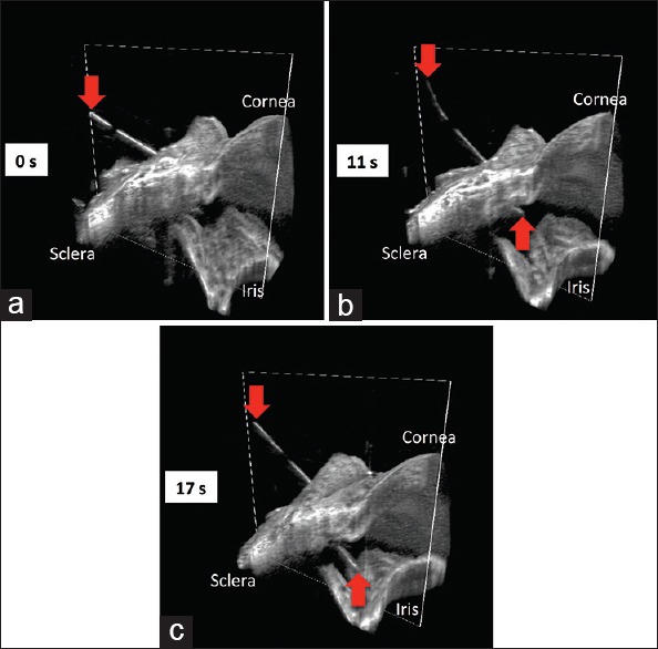
Figure 1.

Swept-source microscope-integrated optical coherence tomography four-dimensional (three-dimensional across time) sequence during scleral tunneling. The 23-gauge needle (red arrows) advancing from the surface of the sclera (a), through the sclera into the anterior chamber (b), and to its deepest point in the anterior chamber (c), creating a scleral tunnel for subsequent tube shunt placement
