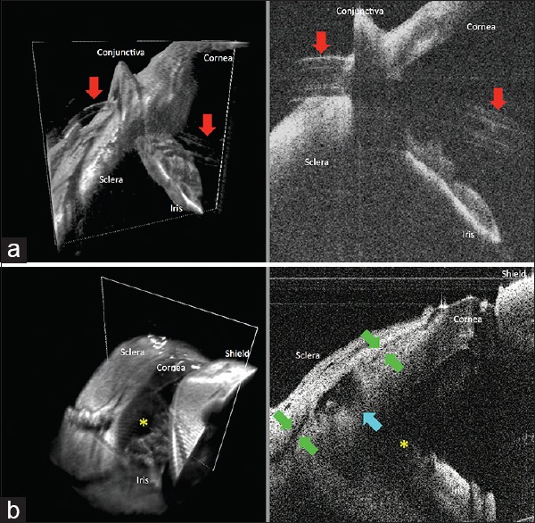
Figure 2.

Swept-source microscope-integrated optical coherence tomography three-dimensional volumes (left) with a white box demarcating its corresponding two-dimensional B-scan (right) at the end of tube shunt placement and trabeculectomy surgeries. (a) Tube shunt (red arrows) insertion through the previously established scleral tunnel into the anterior chamber, with proper positioning anterior to the iris and without corneal touch. (b) Scleral flap interface (green arrows), sclerotomy (blue arrow), and iridectomy (yellow asterisk) at the end of a trabeculectomy surgery
