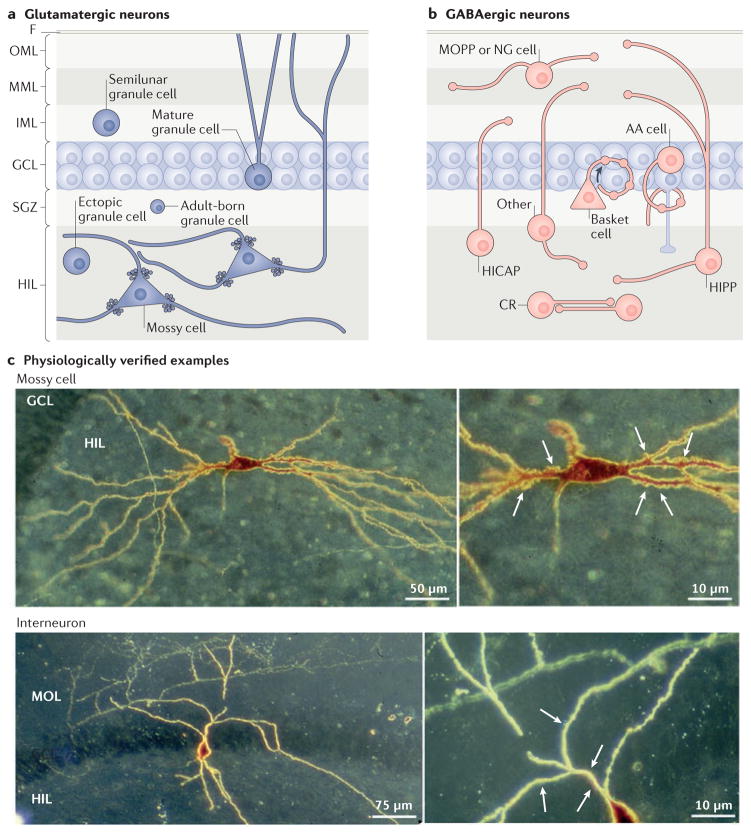
Figure 2. The cell types of the dentate gyrus.
a | Glutamatergic cells of the dentate gyrus include granule cells and mossy cells. Granule cells are not only located in the granule cell layer (GCL); there are small subsets in the inner molecular layer (IML) and hilus (HIL), and precursors to granule cells are located in the subgranular zone (SGZ). Mossy cells have long dendrites, some of which extend into the molecular layer (MOL; comprised of the IML, middle molecular layer (MML) and outer molecular layer (OML))27,28. b | GABAergic neurons of the dentate gyrus are heterogeneous. Their nomenclature is based on the location of the cell body and the axon terminal field13–15. For example, MOPP cells have a cell body in the MOL and terminals in the OML and MML, where the terminals of the perforant path are located13–15. HICAP cells (interneurons with a hilar cell body with axon targeting the commissural/associational pathway) innervate the IML, where the commissural/associational projection from mossy cells is located13–15. The neurons that innervate the granule cell somata or the axon initial segments are called perisomatic-targeting cells. Two of the most common cell types in this group are basket cells, which make basket-like endings around the granule cells and often have a pyramidal-shaped soma that is located at the border of the GCL and HIL134, and axo-axonic (AA) cells13–15. AA cells are often present near or in the GCL, as shown, and innervate granule cell axon initial segments13–15. Several GABAergic neuron subtypes innervate granule cell dendrites. The most common of these are cells in the HIL that innervate the OML and MML (HIPP cells, hilar cells that project to the terminal zone of the perforant path). An example of a neurogliaform cell (NG) that innervates the molecular layer is shown135. There are some types of GABAergic neurons that have an axon that innervates more than one layer (here labelled ‘other’)16,17, and some interneurons innervate each other, such as calretinin-expressing hilar cells (CR)136. c | The upper image shows an example of a biocytin-filled mossy cell with thorny excrescences labelled in the higher magnification inset (indicated by the arrows). The lower image shows an interneuron that was filled with biocytin in a rat hippocampal slice. Arrows point to primary dendrites that are smooth relative to the mossy cell. F, fissure.

