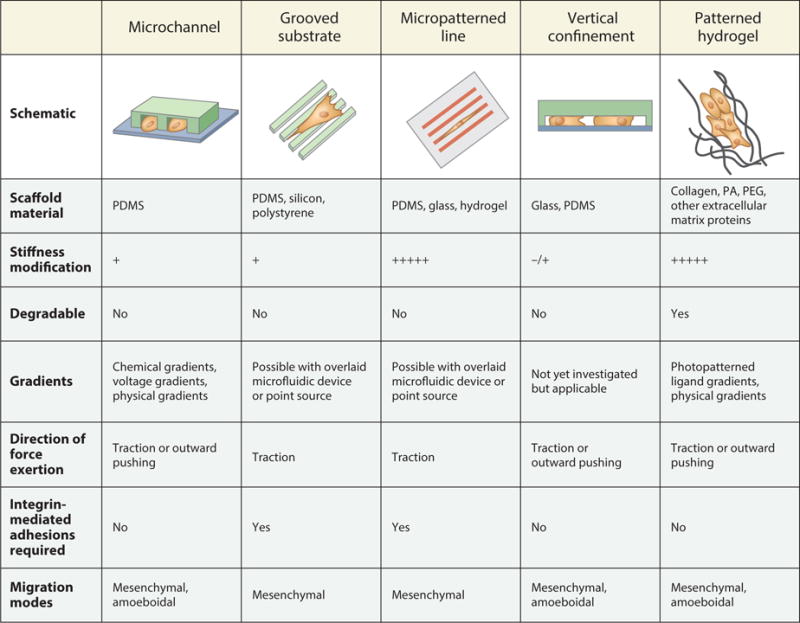
Figure 1.

Schematics of engineered models of cell migration, along with their key physical, mechanical, and mechanistic characteristics. Microfluidic microchannel devices, grooved substrates, micropatterned lines, vertical confinement devices, and patterned hydrogels can be engineered to study cells’ response to confinement. Scaffolds for confined cell migration can be formed from various degradable and nondegradable materials [including polydimethylsiloxane (PDMS), polyacrylamide (PA), and polyethylene glycol (PEG)] with tunable stiffness. Additionally, these scaffolds can be combined with gradients to simultaneously impart multiple cues to migrating cells. Use of these assays has revealed microenvironment-dependent patterns of force exertion and requirements for integrin-mediated adhesions. Importantly, the physical and chemical properties of the model can drive cells to migrate via mesenchymal or amoeboidal mechanisms.
