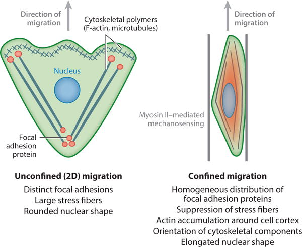
Figure 2.

Schematics of cell morphology in unconfined versus confined microenvironments. Cells migrating on planar, two-dimensional (2D) surfaces form large focal adhesions and stress fibers, have rounded nuclei, and often migrate with a broad leading edge containing branching F-actin structures. Conversely, cells in confinement are characterized by a suppression of large focal adhesions and stress fibers. Instead, focal adhesion proteins are often distributed homogeneously around the cell body. Confined cells typically display alignment of cytoskeletal structures and nuclear elongation along the confining axis.
