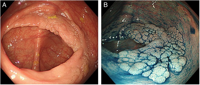
Figure 1.
Forty-millimetre granular-type laterally spreading tumour in the caecum from a patient referred after diagnostic colonoscopy at another centre. (A) High-definition (CF-H290DL, Olympus, Tokyo, Japan) white-light image. (B) After dye spray with indigo carmine chromoendoscopy. This lesion was resected with piecemeal endoscopic mucosal resection (EMR) and histology showed a tubulovillous adenoma with low-grade dysplasia. Images were provided by Dr Malcolm Tan, Translational Gastroenterology Unit, Oxford/Changi General Hospital, Singapore.

